
Estimation of kidneys and urinary bladder doses based on the region of interest in 18fluorine-fluorodeoxyglucose positron emission tomography/computed tomography examination: a preliminary study
Introduction
The combined positron emission tomography (PET) and computed tomography (CT) or known as PET/CT scan has been established as a powerful imaging modality to acquire fusion images of anatomic details and metabolic activity of tissues in a single examination. 18fluorine-fluorodeoxyglucose (18F-FDG) is a glucose analogue and it has been considered as the most frequently used radiotracer for oncology imaging (1). As 18F-FDG is injected into the body, it accumulates in the tissue that has higher glucose level such as tumor cells which will show greater intensity (hot spots) in the PET/CT images.
This high 18F-FDG intensity is not only seen in the tumor cells but also in the normal tissues including kidneys and urinary bladder as 18F-FDG cannot be reabsorbed in the proximal tubule of the kidneys and will be directly excreted through urine into the urinary bladder. Due to the kidney excretion of 18F-FDG, kidney diseases such as lymphoma, leukemia, or metastatic disease can be neglected (2). In addition, the accumulation of 18F-FDG in these organs exposes them to ionizing radiation which can lead to increasing internal radiation dose to the patient. Thus, it is essential to calculate the internal dose to organs from the distribution of radiotracer in both diagnostic and therapeutic nuclear medicine examinations (3).
Since kidneys have complex structural and functional properties, the concern of kidneys and urinary bladder doses in 18F-FDG PET/CT examination is an interesting field of radiation protection which needs more investigations. Hence, precise estimation of radiation dose to the organs concerning the administration of 18F-FDG as a radiotracer is vital in the clinical area. The internal dose to organs from PET is calculated according to the injected radiotracer activity with the use of dose coefficients suggested by ICRP. However, this dose calculation method is not directly measured from the organs; rather it is calculated based on total injected activity of the radiotracer.
The aim of this preliminary study was to estimate the 18F-FDG dose to the kidneys and urinary bladder in whole body PET/CT examination based on region of interest (ROI); assigned as measured dose. The values were then compared to calculated dose from total injected 18F-FDG activity with the application of dose coefficient recommended by the ICRP reports; assigned as calculated dose.
Methods
Participant characteristics
Nine subjects within the age of 19 to 25 y (mean ± SD, 20.78±1.99 y), height from 150 to 172 cm (mean ± SD, 161.11±8.60 cm), and weight from 47 to 74 kg (mean ± SD, 55.44±10.68 kg) were recruited in the whole body PET/CT examination. Table 1 shows a demographic overview of all patients who underwent the PET/CT examination. They fasted for at least six hours and emptied their urinary bladders prior to the examination. Whole body PET/CT scans were performed for 62–70 minutes (mean ± SD, 66±5.66 minutes) after intravenous injection of the 18F-FDG. The injected activity was 262.33–324.12 MBq (mean ± SD, 292.42±20.89 MBq). The study was accepted by the Research Ethical Committee Universiti Teknologi MARA (UiTM): 600-IRMI (5/1/6) as it is related to human as a subject. Each subject had been notified about the research purposes and written informed consent was obtained before they participated in this study. Their involvement was voluntary.

Full table
PET/CT scanning protocol and process
This study used the Siemens Somatom Biograph 64 TruePoint PET/CT scanner (Siemens Medical Solutions, Erlangen, Germany). The subjects were kept lying down in a supine position on the table during the procedure. A CT scout view was then acquired from the vertex to the mid-thigh area for anatomical localization and attenuation correction. A PET image was obtained in a three-dimensional (3D) mode at 2 minutes per bed position. Each PET/CT examination takes about 17 minutes to be completed including 5 minutes to obtain CT image while another 12 minutes for PET scanning.
Calculation of internal organ doses
Internal doses of the kidneys and urinary bladder from 18F-FDG PET scanning were estimated by using two methods which are total injected activity of 18F-FDG (calculated dose) and activity concentration measured within ROI of the organs (measured dose). These two values were then multiplied with recommended dose coefficients for 18F-FDG described in the ICRP 80 (4) and ICRP 106 (5) to estimate organ dose. Thus, two equations were used in this study to estimate organ doses. The first method was calculated using the methodology described in the previous research (6) by the equation:
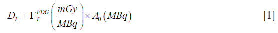
where T is an organ or tissues, DT is the absorbed dose to a kidney or urinary bladder obtained from PET scan, the dose coefficient of kidneys or urinary bladder,  as suggested in Publication 80 and 106 of the ICRP, and A0 is the total injected 18F-FDG activity that was recorded prior to scanning.
The second method was measured using organ activity based on drawn ROI by the following equation:
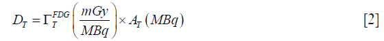
where AT is the kidney or urinary bladder activity in MBq. AT was derived using the methodology described in (7) by the following equation:
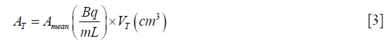
where Amean is the average kidney or urinary bladder activity concentration that was measured from the static whole body 3-D PET image by manually drawing the ROI within each organ across all image planes that contained them. VT is the volume of organ that was measured from CT images based on volume measuring application available on a workstation (Leonardo, Siemens Medical Solutions, Erlangen, Germany). ROI was drawn within the kidneys and urinary bladder in axial CT images which were acquired at 5-mm slice thickness. These drawn ROIs from all slices of axial CT images were automatically added by using the reconstruction interval at the workstation to calculate the organ volume (8).
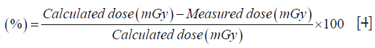
This study assumed direct 18F-FDG uptake with no biological elimination (9). Organ doses were estimated and compared from the two methods of measured organ activity based on ICRP 80 and ICRP 106. Both doses were compared to assess the mean percentage difference between them. The percentage difference between calculated dose and measured dose was calculated by the following equation:
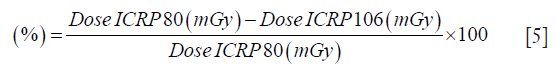
The percentage difference of kidney and urinary bladder doses between ICRP 80 and ICRP 106 was calculated by the following equation:
Results
Organs volume and activity
The volume and activity of the kidneys and urinary bladder were measured from the static whole body 18F-FDG PET/CT images as shown in Table 2. The mean volume of the male left kidney was 150.86 cm3 and the right kidney was 157.16 cm3. While the mean volume of the female left kidney was 126.66 cm3 and the right kidney was 115.51 cm3.

Full table
Kidney doses
The mean kidney doses based on ICRP 80 and ICRP 106 for both genders are shown in Tables 3 and 4, respectively. The mean calculated dose to the male kidneys was slightly lower than the female kidneys. Whereas, no significant difference was observed between the male measured dose for both sides of the male and female kidneys (P>0.05). The mean percentage difference between calculated dose and measured dose to the kidneys ranged from 98.95% to 99.29% based on ICRP 80 and 98.96% to 99.32% based on ICRP 106. This is due to the changes of the dose coefficient for the kidneys from 0.021 to 0.017, which has reduced the absorbed dose to the kidneys. The mean percentage difference of calculated dose to the kidneys between ICRP 80 and ICRP 106 was 19.05% and measured dose ranged from 17.00% to 40.00% as demonstrated in Table 5.

Full table

Full table

Full table
Urinary bladder doses
The mean urinary bladder doses based on ICRP 80 and ICRP 106 are shown in Tables 6 and 7, respectively. The mean calculated dose to the male urinary bladder was slightly lower than the female urinary bladder. While the mean measured dose to the urinary bladder of the males was slightly higher than the female urinary bladder. The mean percentage difference between calculated dose and measured dose to the urinary bladder was 97.08% and 97.27% based on ICRP 80. Whereas, the mean percentage difference was 96.99% and 97.28% based on ICRP 106. The change of the dose coefficient for the urinary bladder from 0.16 to 0.13 had resulted in a decrease in the urinary bladder dose. The mean percentage difference of calculated dose to the urinary bladder between ICRP 80 and ICRP 106 was 18.75% and measured dose ranged from 18.46% to 18.52% as demonstrated in Table 8.

Full table

Full table

Full table
Discussion
With the increasing frequency of whole body PET/CT examination, accurate measurement of the 18F-FDG dose is important as it directly exposes radiosensitive organs. There are several studies that estimate internal dose from administered 18F-FDG (1,6,10,11). However, only one study (1) that estimated the 18F-FDG dose based on activity concentration of organ measured by drawn ROI for both genders. In this current study, kidneys and urinary bladder doses were estimated using two calculation methods which are the total injected activity of 18F-FDG and activity concentration of organ measured by drawn ROI. The measured dose from organs activity concentration was compared with calculated dose from total injected 18F-FDG activity.
A large difference was observed for kidneys and urinary bladder internal doses from administered 18F-FDG between calculated dose and measured dose. The measured dose was less than the calculated dose established in both ICRP 80 and ICRP 106. According to the previous studies (12,13), they reported that the physiological biodistribution of 18F-FDG in the kidneys was 1.3% of the injected dose for the normal human body. Therefore, this current study found that the variability in the distribution of 18F-FDG concentration in the organs may affect the calculation of organ dose based on drawn ROI. This is due to several reasons such as manually drawn ROI for 18F-FDG concentration measurement, the limitation of the ROI size and position and the administered radiotracer activity (1).
The mean calculated dose to the kidneys based on ICRP 80 was slightly higher than a previous study (6). They reported that the PET dose to the kidney was 4.1 mSv which was similar to the absorbed dose to other organs such as the brain, spleen, pancreas, and liver. This is due to the difference in the dose coefficient used for 18F-FDG established by ICRP 80 as they multiplied the dose coefficient value of remaining organs instead of kidneys value with the total injected 18F-FDG activity. Moreover, the mean calculated dose to the urinary bladder in ICRP 80 was higher than the kidneys dose. This result is in agreement with previous studies (6,11), where they reported that the urinary bladder had the highest PET dose; 59.2 mSv, as compared to the other organs with 370 MBq of injected 18F-FDG activity. This is due to the fact that the urinary bladder is considered as the final location of 18F-FDG accumulation as 18F-FDG was excreted through the kidneys.
This study found that there was a reduction in estimated organ doses from ICRP 106 dose coefficient compared to ICRP 80 for 18F-FDG substance. The mean percentage difference between ICRP 106 and ICRP 80 in the male and female kidneys doses ranged from 17.00% to 40.00%, while for urinary bladder doses ranged from 18.46% to 18.52%. A former study (14) stated that effective dose estimation between the ICRP 60 and ICRP 103 tissue-weighting factors showed large differences by 21% to 31% for CT procedures. Hence, the finding in the present study demonstrated that the estimation of organ dose based on ICRP dose coefficient involving administration of the 18F-FDG substance is significant for the future research especially on radiation protection area as it affects the calculation of organ absorbed dose.
The mean calculated dose to the female kidneys was slightly higher than male kidneys according to both ICRP 80 and ICRP 106. Similar to current finding, the mean 18F-FDG dose to the female and male kidneys was recently reported (10) at 5.3 and 4.5 mGy respectively for standard PET/CT examination. While for diagnostic PET/CT examination, the mean kidneys dose for the female was 5.2 mGy and was 4.4 mGy for the male. This may be due to the difference in the administered activity of 18F-FDG for each subject that may affect the calculated dose to the kidneys of males and females. The amount of administered activity and patient’s size are several factors that contribute to the patient radiation dose from PET/CT examination (7).
Inversely, the difference in the mean measured dose between the male and female kidneys was not significant indicating that there was no difference in the distribution of 18F-FDG concentration in the male kidneys and female kidneys. This finding is in agreement with the previous study (7), in which 18F-FDG biodistribution in the male and female brains were reported to have no difference due to the fact that there is no significant difference in the residence times of 18F-FDG activity. They assumed that the 18F-FDG distribution in the brain depends on the selection of subject’s characteristics for the research. The distribution of 18F-FDG in the male and female varied because of physiological differences (7,15), between genders.
The inconsistency in measured dose to the urinary bladder in this study can be observed due to the difference in the urinary bladder activity and volume, where the highest was 27.94 MBq. According to previous studies (16,17), they assumed that the differences in the reabsorption of 18F-FDG by renal tubule could affect the activity of the urinary bladder. The urinary bladder has an elastic wall where the volume depends on the volume of the contained urine (18). Thus, an underestimation of measured dose to the urinary bladder would occur if the measurement of urinary bladder dose is estimated before 18F-FDG injection.
Conclusions
This study demonstrated that the estimation of organ 18F-FDG activity based on drawn ROI and the use of ICRP 106 dose coefficient results in a decrement of organ absorbed dose for both genders. Therefore, the application of new organ activity estimation and the latest version of ICRP 106 dose coefficient should be further explored by research committees in order to produce accurate internal dose received by a patient in the whole body PET/CT examination.
Acknowledgements
The authors would like to thank Mr. Rosli and Mr. Amirul for helping during the scanning process and providing suggestions.
Footnote
Conflicts of Interest: The authors have no conflicts of interest to declare.
Ethical Statement: This study was supported by Faculty of Health Sciences, Universiti Teknologi MARA (UiTM) Puncak Alam Campus under Research Acculturation Grant Scheme (RAGS): 600-RMI/RAGS 5/3 (150/2014). Each subject had been notified about the research purposes and written informed consent was obtained before they participated in this study.
References
- Khamwan K, Krisanachinda A, Pasawang P. The determination of patient dose from 18F-FDG PET/CT examination. Radiat Prot Dosimetry 2010;141:50-5. [Crossref] [PubMed]
- Zukotynski K, Lewis A, O’Regan K, Jacene H, Sakellis C, Almodovar S, Israel D. PET/CT and renal pathology: a blind spot for radiologists? Part 2-lymphoma, leukemia, and metastatic disease. AJR Am J Roentgenol 2012;199:W168-74. [PubMed]
- Laffon E, Bardiès M, Barbet J, Marthan R. Calculating an estimate of tissue integrated activity in 18F-FDG PET imaging using one SUV value. EJNMMI Res 2013;3:26. [Crossref] [PubMed]
- ICRP. Radiation dose to patients from radiopharmaceuticals (addendum 2 to ICRP Publication 53). ICRP Publication 80. Ann ICRP 1998;28:1-126. [Crossref]
- ICRP. Radiation dose to patients from radiopharmaceuticals (a third amendment to ICRP Publication 53). ICRP Publication 106. Ann ICRP 2008;38:1-197. [PubMed]
- Huang B, Law MW, Khong PL. Whole-body PET/CT scanning: estimation of radiation dose and cancer risk. Radiology 2009;251:166-74. [Crossref] [PubMed]
- Kaushik A, Jaimini A, Tripathi M, D'Souza M, Sharma R, Mondal A, Mishra AK, Dwarakanath BS. Estimation of radiation dose to patients from 18FDG whole body PET/CT investigations using dynamic PET scan protocol. Indian J Med Res 2015.721-31. [PubMed]
- Shin HS, Chung BH, Lee SE, Kim WJ, Ha H. Il, Yang CW. Measurement of kidney volume with multi-detector computed tomography scanning in young korean. Yonsei Med J 2009;50:262-5. [Crossref] [PubMed]
- Zanotti-Fregonara P, Jan S, Taieb D, Cammilleri S, Trebossen R, Hindie E, Mundler O. Absorbed 18F-FDG dose to the fetus during early pregnancy. J Nucl Med 2010;51:803-5. [Crossref] [PubMed]
- Quinn B, Dauer Z, Pandit-taskar N, Schoder H, Dauer LT. Radiation dosimetry of 18F-FDG PET/CT : incorporating exam-specific parameters in dose estimates. BMC Med Imaging 2016;16:4-1. [Crossref] [PubMed]
- Brix G, Lechel U, Glatting G, Ziegler SI, Münzing W, Müller SP, Beyer T. Radiation exposure of patients undergoing whole-body dual-modality 18F-FDG PET/CT examinations. J Nucl Med 2005;46:608-13. [PubMed]
- Zincirkeser S, Sahin E, Halac M, Sager S. Standardized uptake values of normal organs on 18F-fluorodeoxyglucose positron emission tomography and computed tomography imaging. J Int Med Res 2007;35:231-6. [Crossref] [PubMed]
- Yasuda S, Takahashi W, Takagi S, Fujii H, Ide M, Shohtsu A. Factors influencing physiological FDG uptake in the intestine. Tokai J Exp Clin Med 1998;23:241-4. [PubMed]
- von Boetticher H, Lachmund J, Looe HK, Hoffmann W, Poppe B. 2007 recommendations of the ICRP change basis for estimation of the effective dose : what is the impact on radiation dose assessment of patient and personnel? Rofo 2008;180:391-5. [Crossref] [PubMed]
- Kaushik A, Jaimini A, Tripathi M, D’Souza M, Sharma R, Mishra AK, Mondal A, Dwarakanath BS. Estimation of patient dose in 18F-FDG and 18F-FDOPA PET/CT examinations. J Cancer Res Ther 2013;9:477-83. [Crossref] [PubMed]
- Mejia AA, Nakamura T, Masatoshi I, Hatazawa J, Masaki M, Watanuki S. Estimation of absorbed doses in humans due to intravenous administration of fluorine- 18- fluorodeoxyglucose in PET studies. J Nucl Med 1991;32:699-706. [PubMed]
- Woosley RL, Kim YS, Huang KC. Renal tubular transport of 2-Deoxy-D-Glucose in dogs and rats. J Pharmacol Exp Ther 1970;173:13-20. [PubMed]
- Viswanathan AN, Yorke ED, Marks LB, Eifel PJ, Shipley WU. Radiation dose-volume effects of the urinary bladder. Int J Radiat Oncol Biol Phys 2010;76:S116-122. [Crossref] [PubMed]

